Below you can find some samples of the EIBR research:
Brain Samples
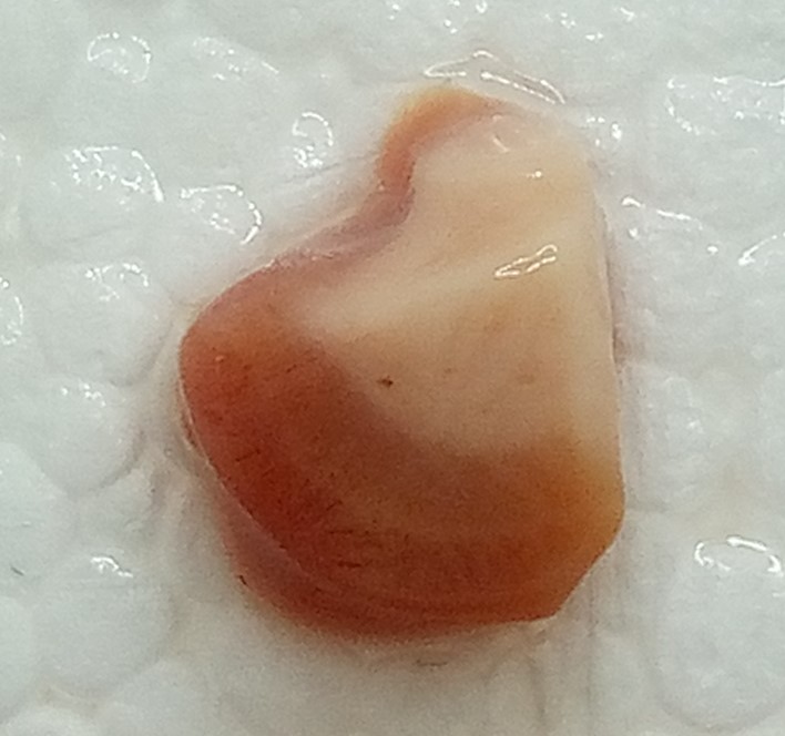
We obtained several high-quality human brain samples in the Netherlands. We are very grateful for the people who were willing to donate their bodies for this research. The picture shows a piece of the human brain from the primary visual cortex. The darker part of the chunk of tissue in the picture, on the bottom, closer to the surface of the cortex, is where the cell bodies of neurons are (gray matter). The part on the top is lighter (white matter) because what you can find there is mostly the parts of the neurons called axons, which are like wires that go to other neurons. The whiteness comes from supporting cells (glia), which wrap around the axons like insulation around electrical wires. Such a coating (called myelin) allows the electrical impulses to travel quickly. But if you look closely, you can also see a white stripe parallel to the surface of the cortex. Such a stripe is visible only in the primary visual cortex of primates (a group of animals that includes monkeys).
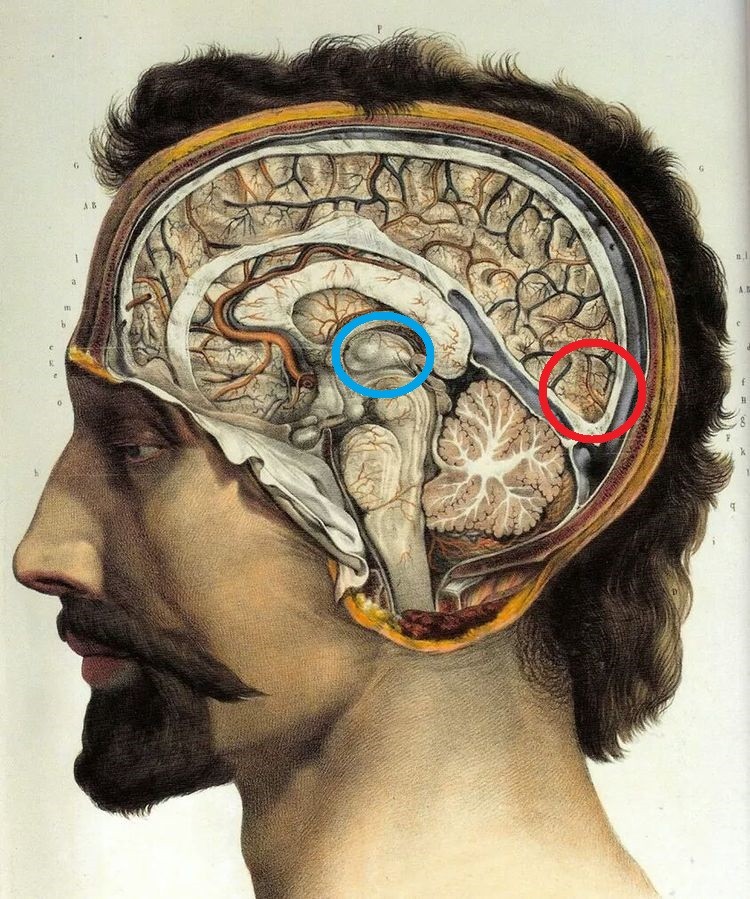
If you feel the back of your head, you can feel a small bump. Roughly below this bump is, on each hemisphere of the brain, is the part of the cortex that is the first to process the visual information (red circle in the picture). The neurons that receive this information lie in a specific layer of the cortex (layer 4). But they do not connect directly to the eye. Rather, they connect to neurons in a structure deep in the brain, called the thalamus (blue circle in the picture). For primates, quick processing of visual information is very important. A white line is present in the primate primary visual cortex because a lot of axons—insulated with myelin and originating in the thalamus—end there.
Autapses enable pattern recognition in neural networks
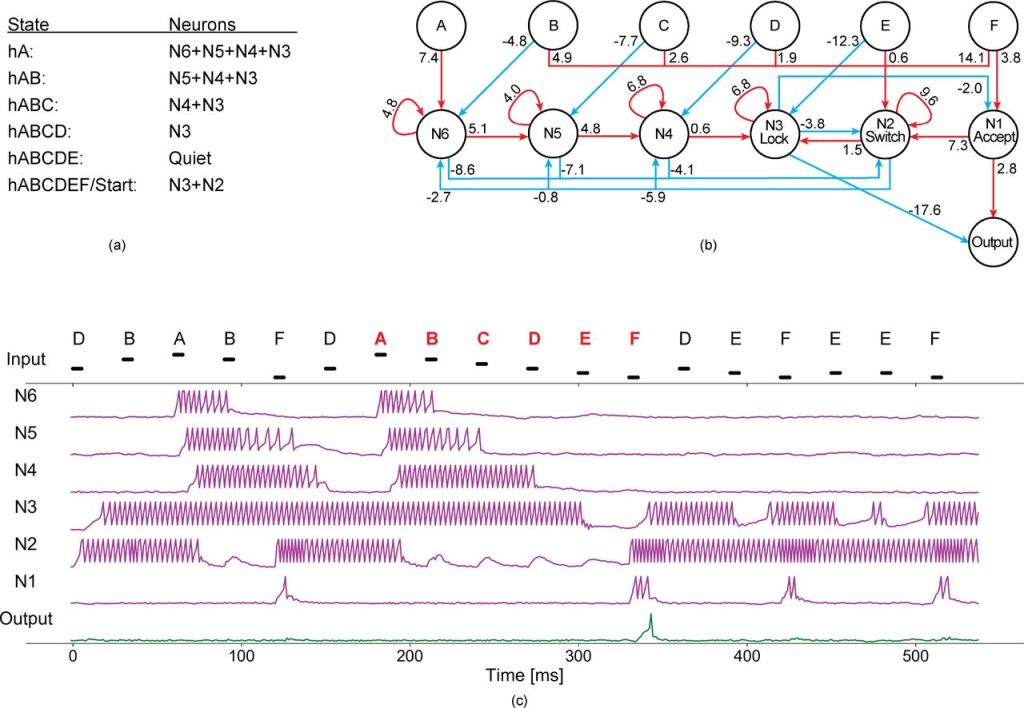
In this EIBR paper we show how autapses (a connection of a neurone onto itself) are important for maintaining network state, a form of short-term memory. The picture below shows a network of artificial neurons recognizing a simple pattern consisting of 6 input signals. When the network receives the signals in a specific order (symbols in red), the output neuron (whose membrane voltage is shown in green at the bottom) becomes active (the voltage increases and falls down again, this is called a spike).
If you want to read more, have a look at https://www.biorxiv.org/content/10.1101/2023.11.16.567361v1.full
